Fibromyalgia
Patient MM was referred from her physio for non-resolving left anterior hip and iliac crest pain with standing and functional muscle pain in the left calf and stiffness of the left anterior ankle. Learn how Formthotics got her back to doing what she loved.
Presenting Complaint
Referred from her physio for non-resolving left anterior hip and iliac crest pain with standing and functional muscle pain in the left calf and stiffness of the left anterior ankle.
History of the presenting complaint
The patient gets sustained discomfort and hip stiffness with any periods of extended walking or standing with NDA (Normal Daily Activities). This has been present for 12+ months. She has been receiving physio treatment, consisting of hip and pelvic mobilisation and fascial release of Iliopsoas, Rectus Femoris and Adductors. This has provided some short-term relief, post-treatments, but no lasting resolution. She had also been prescribed calf raises, but these increased her calf pain, especially in the left calf.
Past Medical History
Patient MM has a history of fibromyalgia with a diagnosis 20 years ago. She has been an active walker/tramper. She does continue walking on a regular basis as she needs this for life balance and general fitness.
She has had various fibromyalgic symptoms over the last 20+ years, mainly centred on the lower lumber back/hips and, of late, her left posterior calf and ankle.
Assessment
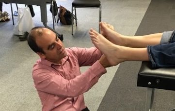
Base vascular and neural tests were unremarkable. Reflexes present.
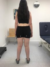
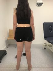
On static stance, there was a genu valgum (knock knees), greater on the left. Lateral position of right patella noted. Left side transverse anterior pelvic rotation. Left iliac crest was positioned lower, as was the left gluteal cleft. Slight thoracic curvature to left. RCSP (Resting Calcaneal Stance
Position) valgum (eversion) on left. Left foot navicular drop exceeded drift, and Supination Resistance Test (SRT) was high, with the 1st MTPJ being dorsiflexed, leading to a possible Functional Hallux Limitus.
The right side MLA (Medial Longitudinal Arch) profile was higher with an easier SRT (Supination Resistance Test). Jack’s test was adequate on the right only. A small amount of metatarsus abduction was seen on the right foot.
She had greater instability on the left leg with single-leg balance, but adding movement over this displayed bilateral medial knee drift, driven proximally from the hip. Single leg heel raises could be performed with a high heelhold, but she fatigued earlier on the left.
Knee to wall lunge test showed she had adequate distance when she compensated by dropping and driving the hip on the affected side, forward, but when instructed on correct positioning, she had 3cm on the left side and 5cm on the right. When we had Patient MM non-weight-bearing on the plinth, at rest, the left leg sat externally rotated. There was no discernible structural leg length disparity with umbilicus to malleoli measurement or skyline knee measurements. The right SIJ (Sacroiliac Joint) rotation test showed a deficit of internal rotation, and there was good ROM (Range of Motion) in both hips.
Straight leg raise was less than 90 degrees, bilaterally, and had greater neural tightness in the left leg.
Manual Resisted Activation and Resistance Tests were a little more interesting. On the left side, Tibialis Anterior and Extensor Digitorum Longus (EDL) had poor resisted strength, while Peroneus Longus activation was unachievable for her.
On the Right side, the Tibialis Posterior had poor activation and EDL on this side poor resisted strength. Straight leg adduction was adequate while she struggled with resisted abduction. Gluteus Medius bilaterally had poor activation, as did the left hamstring group. Prone hip flexor tightness was noted on the left side.
Bilaterally, she had adequate ROM in the 1st MTPJ, but there was marked hypomobility in the lateral column on the left foot, especially in relation to cuboid plantar glide. Both superior tibiofibular joints had poor anterior glide. Passive, active, and forced ankle joint ROM all had limitations in dorsiflexion on the left side.
With both feet, she had poor elicitation of intrinsic function with short arch pull, digital adduction/abduction, etc.
Observing her walking, the right foot had increased abductory twist at heel lift. There was also a greater arm swing on the right side.
Left foot contact was further lateral of the mid-line. At mid-stance, on the left side, she made contact in an everted position moving through with a medial loading.
Working with this, we felt that she had an underlying functional length discrepancy and had compensatory patterning around this.
Our starting point was block testing for height-differential based on marked positions. The medial column loading on the left foot we felt was exacerbated by increased femoral angle and pelvic drop.
Patient Goals
Patient MM goals were to reduce pain in the lower lumbar back and hip with normal daily activity and to be able to walk for 2+ hours pain-free.
Treatment Plan
She wears a low-pitched shoe, as these are the most comfortable for her, and she does not wish to change these.
The decision was made to fit Original Single Medium Formthotics and heat-mould them in the shoes. On the left foot, we Initially added a 4mm heel raise, extended to the midfoot. A small medial rearfoot skive was applied bilaterally to exert a supinatory force, medial to the marked STJ (Subtalar Joint) axial line. The heel was balanced laterally to reduce lateral spill. A small 2mm extended PMP (Proximal Metatarsal Phalangeal Pad) was applied to aid sagittal movement.
We initiated a course of mobilisation and manipulation to alter accessory joint movement and increase ankle joint ROM. This would be reviewed and continued over a 4 to 6-week period.
Added to this was some functional patterning exercises for ankle stability based around engagement of ankle extensor and evertors. Mainly these were tripod holds with excursions.
She would continue hip strengthening as per physiotherapist instructions.
Patient MM was also asked to trial a period without wearing the modified Formthotics to see if there was any perceivable change +/-.
Outcome
Upon first review, ten days after the initial assessment, MM noted that she indeed felt that her pain was improving but still present after an extended time on her feet. Her feeling was that she was now not high enough with the adjustment we had made, and when she did not wear the Formthotics, she noticed the imbalance.
Her functional strength had improved, and she remained diligent at her exercises.
The ankle initially felt as if it had better movement and was not “jamming” but slowly reverted after about a week. On exam, decreased Talocrural posterior glide was noted.
From here, we made a small incremental height adjustment, continued the course of mobilisations/manipulations, added a kneeling ankle stretch.
She had four return visits for manual therapy and orthotic review. Within three months, she returned to tramping in the hills with minimal back/hip and leg discomfort.
She has continued wearing the orthoses and is comfortable at present with the modifications.
Ongoing, the plan is to increase her walking/tramping, and we will review overall foot/ankle strength and movement parameters every three months.